In-Hospital Gastroenterology Procedures in Columbus, GA
If diagnosis or treatment for your gastrointestinal condition requires a procedure performed in the hospital, rely on the gastroenterologists at Southeastern Gastroenterology. Our GI specialists are available to provide exceptional care to patients in West Central Georgia or East Central Alabama.
Southeastern Gastroenterology performs in-hospital tests, exams, and procedures for the most comprehensive GI care possible.
If you are scheduled to undergo a procedure, please view our prep instructions for all procedures.
settings
Endoscopic Procedures
settingsAn anal manometry test, also called anorectal manometry, is a test that evaluates the anus and rectum’s pressure and muscle function. It particularly evaluates the function of the anorectal sphincter, or the muscles that control when stools are released. Patients who suffer from fecal incontinence, constipation, or bowel movement difficulties may require anorectal manometry to determine the cause of their issue(s).
What does anorectal manometry entail?
Your specialist will insert a catheter with pressure sensors into the rectum. You may be asked to squeeze, tighten, or push your rectal muscles to evaluate strength and function. This exam can last 30-90 minutes. While typically painless, this test may feel uncomfortable for the duration, and it may cause you to feel as if you are having a bowel movement.
What happens after anorectal manometry?
This exam does not require anesthesia or sedation of any kind. You are free to drive yourself home after. Your specialist will have results within one to two weeks following the exam.
A colonoscopy is a crucial procedure that evaluates the health of the colon and checks for colon polyps, colon cancer, and other changes or abnormalities.
It’s important for men and women 45 years or older, as well as younger patients at risk of colon cancer, to undergo routine colonoscopies every 10 years, or more frequently if directed by their doctors.
What does a colonoscopy entail?
A colonoscopy is not a difficult or complex procedure, but it does require some preparation before it can be performed.
- Preparation: Your GI doctor will give you specific instructions to prepare your bowels for the procedure. On the day before your procedure, you will start a clear liquid diet (water, broth, clear sodas, white grape juice, tea, and coffee without cream or milk, etc.) and avoid solid foods of any kind. You will also take a prescribed laxative to clear out your bowels completely. Follow all instructions carefully.
- Procedure: At your procedure, you receive some amount of sedation for a more comfortable experience. You will lie on your side of the examination table. Your doctor will insert a colonoscope into the rectum. Air or carbon dioxide may be pumped into the rectum to inflate it for a better look at your colon’s wall and structures. Your doctor will look for signs of trouble, including polyps, cancer, or other structural issues. If there are polyps, your doctor will take a biopsy or remove the polyps completely. Once completed, the colonoscope is removed, and the procedure is over.
What happens after a colonoscopy?
After your colonoscopy, you will be sent to a recovery room for observation until you are released.
Every colonoscopy patient will require a driver after being released from the hospital.
Endoscopic retrograde cholangiopancreatography (ERCP) is an endoscopic procedure that checks for stones, tumors, or signs of narrowing in a patient’s bile ducts and pancreatic ducts. When a patient experiences issues with their gallbladder or pancreas, it could be a result of duct narrowing or blockage. An ERCP allows GI doctors to evaluate these organs and systems to detect or rule out the cause.
What does an ERCP entail?
Your doctor will insert an endoscope into the mouth, through the stomach, and down to the duodenum, or the first part of the small intestines, to reach the pancreas, gallbladder, and bile ducts. Once at the right areas, they will inject a dye into the ducts and observe its movements through X-ray technology. If there are stones or other blockage, your doctor may be able to complete removal depending on size and location. They may also be able to aid in duct draining or place stents to keep the ducts open.
Patients will need to fast for several hours before the procedure and will receive sedation for a more comfortable experience.
What happens after an ERCP?
You may experience mild bloating or intestinal discomfort after the procedure, which should fade quickly. You will recover in the hospital for some time before being released.
All ERCP patients will require a driver after being released from the hospital.
Some patients who have experienced narrowing in their esophagus may require an esophageal dilation to widen the esophagus and allow for better swallowing again. Whether caused by chronic acid reflux or GERD, injury, a hiatal hernia, or even esophageal scarring from radiation therapy, a narrowed esophagus requires widening for better eating, drinking, and overall well-being.
What does esophageal dilation entail?
This is a minimally invasive procedure that requires your doctor to insert an endoscope into the mouth and down to the narrowed part of the esophagus. Using either a balloon catheter, dilator, or weighted and cone-shaped tube called a bougie, your doctor will inflate the narrowed section with the balloon or pass the dilator or bougie through the narrowed section to gently re-widen the esophageal wall. X-ray imaging is used to help guide the instrument(s) used.
What happens after esophageal dilation?
Patients receive sedation for a more comfortable experience, so they are monitored for a period of time after the procedure before being released.
All esophageal dilation patients will require a driver after being released from the hospital.
An esophageal manometry test, sometimes called an esophageal motility study, measures the pressure, movement, and function of the esophagus and the muscles that control the esophagus. This test identifies weaknesses or problem areas in the esophagus, and it is especially helpful in diagnosing swallowing issues or sources of non-cardiac chest pain.
What does an esophageal manometry test entail?
Your doctor will insert a thin tube with pressure sensors through the nose and into the esophagus. Once in place, you will be asked to swallow several times to evaluate the strength of your esophageal muscles, the way they contract and relax, and more.
Patients will need to fast for several hours before the procedure, but typically, sedation is not used.
What happens after an esophageal manometry test?
Patients are released relatively soon after the test is over and do not require a driver.
Patients suffering from a variety of esophageal conditions or complications may need an esophagogastroduodenoscopy so their doctor can inspect the condition of their upper GI tract.
What does an EGD entail?
This is an endoscopic procedure where a thin, flexible tube, complete with a camera and a light, is inserted into the mouth and through the esophagus. It gives your doctor a first-hand look at your upper digestive tract from the esophagus to the stomach and duodenum, or the first section of the small intestines.
Based on what they observe through the EGD, your GI doctor can better determine the causes or conditions of your esophageal problems or symptoms.
EGD with Bravo Testing
An EGD with Bravo pH testing follows the same steps as a normal EGD. However, it also involves measuring the acid levels in the esophagus. This is especially helpful for diagnosing issues like GERD.
What does an EGD with Bravo pH testing entail?
While the endoscope is inserted for the EGD, your doctor will also place a small capsule onto the lining of the esophagus, which will measure how acidic the esophagus is. The capsule will remain in place for 48-96 hours or until it naturally falls off. While in place, it will send recordings to a receiver, which your doctor will be able to analyze.
While in place, you will be asked to record things like symptoms, activities, foods eaten, and more for your doctor to compare to the data produced during the test.
What happens after an EGD or an EGD with Bravo Testing?
Patients will receive sedation for a more comfortable experience, so they will be monitored for some time after the procedure before being released.
All EGD patients will require a driver after being released from the hospital.
Patients with issues related to the rectum or colon may require a flexible sigmoidoscopy to inspect part of the colon and diagnose conditions. Not quite as involved as a colonoscopy, a sigmoidoscopy only observes the lower part of the large intestine (the sigmoid). A colonoscopy observes the entire colon.
What does a flexible sigmoidoscopy entail?
A flexible sigmoidoscopy is not a difficult or complex procedure, but it does require some preparation first.
- Preparation: Your GI doctor will give you specific instructions to clear your bowels before the procedure. This typically involves some fasting and taking prescribed laxatives according to your doctor’s instructions.
- Procedure: At your procedure, you will lie on your side on the exam table with your knees pulled up to your chest. Your doctor will insert the sigmoidoscope into your rectum and gently guide it to the lower colon (the sigmoid colon). Your doctor will examine the structures of your colon and look for any abnormalities. During a sigmoidoscopy, your doctor may take a biopsy or remove a polyp if necessary. Once completed, the sigmoidoscope is removed, and the procedure is over.
What happens after a sigmoidoscopy?
If sedation is used, you will be required to recover in the hospital for a time before being released, and you will need a driver before being discharged. If sedation is not used, you can drive home shortly after the procedure has ended.
Hemorrhoid banding, sometimes called rubber band ligation, is a procedure to treat and remove bothersome hemorrhoids, or swollen veins found in the lower rectum or anus.
What does hemorrhoid banding entail?
You will likely be asked to lie on your side on the exam table with your knees pulled to your chest. Your doctor will insert an anoscope (a small tube with a light) to access the hemorrhoids. Using a ligator, your doctor will grasp the hemorrhoid and place the rubber band around it at its base. Over time, the rubber band will cut off the blood supply to the hemorrhoid, forcing it to shrink and fall off.
Hemorrhoid banding typically does not involve sedation. You may feel some discomfort, but it should not be a painful procedure.
What happens after hemorrhoid banding? Patients can resume normal activities shortly after the procedure. Since sedation is not used, patients can also drive home.
A PEG (percutaneous endoscopic gastronomy) tube is a valuable tool for helping patients who are unable to eat or drink normally. Through the PEG tube, patients can receive nutrients, medications, water, and other necessary substances effectively and efficiently.
What does PEG tube placement entail?
You will either undergo sedation or receive local anesthesia before the procedure begins. Your GI doctor will use an endoscope to locate the part of the digestive tract for tube placement. Once located, they will make a small incision somewhere on the abdomen to feed the tube through to the stomach. They will also carefully make an incision in the stomach to insert the tube. Small discs and devices are used to hold the tube in place, both in the stomach and on the abdomen walls. Once complete, the endoscope and other tools are removed, and the procedure is over.
What happens after PEG tube placement?
You will recover in a comfortable room following placement until your sedation wears off. You will receive care instructions, as well as instructions for how to use your tube and any components needed for use.
All PEG tube placement patients will require a driver after being released from the hospital.
Pill cam imaging, or a capsule endoscopy, is an alternative to traditional endoscopy procedures. Using a small, capsule-sized camera, your doctor can gather images of your entire GI tract, from the esophagus all the way to the rectum.
Pill cams are most effective at observing the condition of the small intestines, which cannot be reached endoscopically or colonoscopically. Patients suspected of having celiac disease, Crohn’s disease, or other small intestinal issues are likely to require pill cam imaging.
What does pill cam imaging entail?
You will be given a pill cam, complete with a small camera, light, and transmitter, to swallow. Once swallowed, the pill travels through the esophagus, stomach, small intestines, and large intestines until it is ready to exit. This camera sends signals to a receiver to capture images. It is also disposable, so once it is out, there is no need to collect it.
There is no sedation required for pill cam imaging; it is just like swallowing a pill or vitamin.
What happens after pill cam imaging?
Patients are able to leave shortly after consuming the camera and can resume normal activities immediately.
Imaging Tests
settings
A CT (computed tomography) scan is often required to evaluate organ function and internal conditions and diagnose certain gastrointestinal problems. Using X-ray and computer technology, the GI specialists at Southwestern Gastroenterology can evaluate the GI-specific organs and tissues found in the abdomen or abdominal area of the body.
How does a CT scan work?
Patients will lie still on the table of the CT machine while the donut-shaped scanner captures images of the abdomen from several different angles. The computer receives the images and combines them to create cross-sectional images of your organs and internal structures.
CT scans are helpful in detecting tumors, cysts, or other organ abnormalities. They can also reveal injuries and determine the source of symptoms like abdominal pain and other GI-related issues.
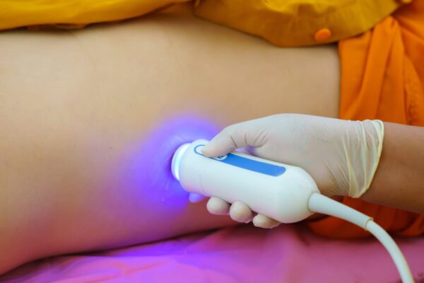
A FibroScan is effective at evaluating the health of the liver. Specifically, it reveals any scarring (fibrosis) or determines if the liver has developed steatosis (become fatty).
How does a FibroScan work?
A FibroScan relies on ultrasound technology to examine the liver. The speed of the soundwaves released through the ultrasound’s transducer can reveal if the liver has developed fibrosis or steatosis. If the soundwaves bounce at a faster pace, it indicates the liver has a more rigid surface due to issues like fibrosis or steatosis. If the soundwaves bounce back at a slower pace, it suggests that the liver is softer, a sign of a healthy liver.
FibroScans are helpful in diagnosing issues like cirrhosis, non-alcoholic liver disease, hepatitis, and more.
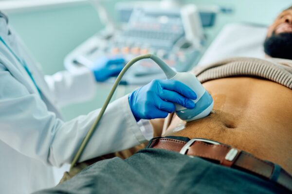 Oftentimes, GI doctors will utilize ultrasound technology along with CT scans or X-ray scans to thoroughly evaluate a patient’s internal structures.
How does an ultrasound work?
Ultrasounds use high-frequency sound waves that are emitted into the body. The echoes that the soundwaves create are interpreted by a computer system and converted into images that are displayed on the monitor, creating real-time images of the patient’s insides.
Ultrasounds can help with diagnosing or evaluating several gastrointestinal issues, including acute abdominal issues, tumor development, cancer development, Crohn’s disease, ulcerative colitis, kidney stones, gallstones, liver conditions, pancreatic conditions, and more.
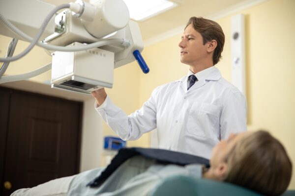
X-rays, particularly fluoroscopic X-rays, can reveal specific abnormalities in the body related to the GI tract and the organs connected to the digestive system. The most common types of GI-related X-rays include upper GI series to evaluate the esophagus, stomach, and upper part of the small intestine (duodenum); lower GI series to evaluate the lower part of the small intestine (ilium), large intestine (colon), and rectum; and abdominal series for a more “bird’s eye view” of the digestive tract.
How does fluoroscopy work?
Traditionally, X-ray technology sends rays of electromagnetic radiation through the body so that harder, more dense tissue, like bones, will absorb the radiation and appear clearly in the images captured by the imaging system. Since the tissues of the organs and structures connected to the GI tract are softer, they require a contrast dye that will appear clearly in the system. Through fluoroscopy, a dye is applied to the observed area and tracked using X-ray imaging. The GI specialist will follow the path of the dye and check for abnormalities that the dye reveals.
Fluoroscopic X-rays can help evaluate or diagnose ulcers, system blockages, polyps, tumors, perforations, and other issues.
Infusion Services
settingsSome GI patients require infusion therapy along with their treatment or management of their condition, especially if they are unable to consume foods and medicines orally or if their GI system does not absorb medications or nutrients properly.
How does Infusion work?
Infusion introduces medications, nutrients, fluids, and other necessary substances into the bloodstream through an intravenous (IV) line. Every patient is different, so some may require infusion treatments more frequently than others.
Commonly, patients with Crohn’s disease or ulcerative colitis require infusion therapy.